An impacted wisdom tooth is not simply a tooth that has failed to erupt: its angle, depth, position against the second molar, and relationship to nerves or the sinus determine the problem and the treatment. By the end, you will know which symptoms need assessment, when removal is justified, what imaging and anaesthesia involve, and how to compare treatment quotations safely.
Key takeaways
- A panoramic radiograph shows the tooth’s angle, depth, roots, and relation to the nerve.
- Monitor an asymptomatic tooth when examination finds no disease or damage and follow-up is reliable.
- Ask how pain control, stitches, eating, brushing, and emergency contact instructions will be handled.
- Seek urgent advice for worsening swelling, fever, trouble swallowing, or bleeding that will not stop.
How to tell whether a wisdom tooth is impacted
An impacted wisdom tooth is blocked from reaching its normal position because of its angle, depth, gum, bone, or the neighbouring second molar. It may cause no symptoms, so an oral examination and panoramic radiograph are the usual starting points.
| Type | What it means | Why it matters |
|---|---|---|
| Mesioangular | The crown tilts forward toward the second molar | It can trap food or damage the second molar |
| Vertical | The tooth is upright but lacks space to erupt | It may remain partly or fully buried |
| Distoangular | The crown tilts backwards toward the jawbone | Removal can be more difficult |
| Horizontal | The tooth lies almost on its side | Its crown may press against the second molar |
| Soft-tissue impaction | Gum covers part of the tooth, but bone does not | The gum flap can trap bacteria and cause pericoronitis |
| Bony impaction | Bone covers part or all of the tooth | Surgical exposure and sectioning may be needed |
| Second-molar impaction | The wisdom tooth is wedged against the tooth in front | Decay, root damage, or periodontal damage can develop |
Ask for assessment if you have:
- Repeated gum swelling, pain, or infection behind the second molar
- A bad taste, pus, or repeated food trapping under a gum flap
- Difficulty opening your mouth or pain at the back of the jaw
- Cheek biting, visible decay, or damage to the second molar
- A cyst or other change found on an X-ray
A CBCT scan is reserved for cases where the inferior alveolar or lingual nerve, adjacent tooth, or jaw structures need three-dimensional assessment. This evaluation helps plan wisdom tooth extraction in Nashik; symptoms alone do not prove that removal is necessary.
When removal is necessary—and when monitoring is safer
Removal is warranted when disease or a clear treatment need outweighs the surgical risk. Monitoring is safer when an impacted tooth is symptom-free, cleanable, and shows no damage on examination or imaging.
Common reasons to recommend removal include:
- Recurrent or severe pericoronitis around a partially erupted lower tooth
- Decay that cannot be restored in the wisdom tooth or second molar
- Periodontal bone loss or a deep pocket beside the second molar
- Cystic change, abscess, root resorption, or another pathological finding
- A tooth that blocks planned orthodontic, prosthetic, or jaw surgery
| Option | What it means | When it applies |
|---|---|---|
| Removal | Surgical extraction, sometimes involving gum incision, bone removal, and tooth sectioning | Disease, repeated infection, non-restorable decay, or a defined treatment need |
| Monitoring | Examination and periodic imaging, with cleaning advice and review if symptoms develop | No disease, no treatment need, and a riskier operation |
| Modified treatment | Delay surgery, change anaesthesia planning, or consider coronectomy | Medical risk or nerve proximity makes standard removal less suitable |
For a lower tooth close to the inferior alveolar or lingual nerve, discuss altered sensation in the lip, chin, or tongue before consent. Coronectomy may reduce nerve-injury risk by leaving healthy, stable roots, but infected, mobile, or diseased roots usually rule it out.
An upper tooth near the maxillary sinus needs separate discussion: persistent fluid from mouth to nose, nasal discharge through the socket, or a wound that will not close requires prompt review. Ask what the proposed third molar removal in Nashik includes, especially imaging, anaesthesia, medicines, follow-up, and complication management.
What imaging, surgery, and anaesthesia involve
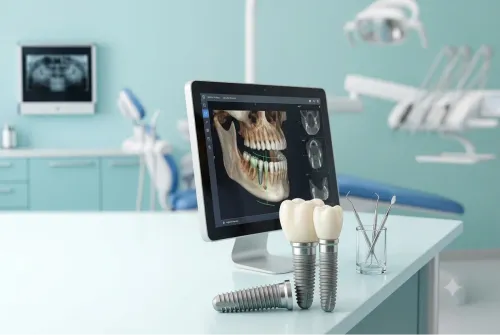
A panoramic radiograph usually maps the tooth’s angle, depth, roots, adjacent second molar, and nearby jaw structures. CBCT adds three-dimensional detail when roots appear close to the inferior alveolar nerve, lingual nerve, maxillary sinus, or another tooth. Examination also checks mouth opening, infection, and bone coverage.
Searching for painless tooth extraction in Nashik should not mean expecting every impacted tooth to be simple or completely sensation-free.
- The clinician numbs the area and makes a gum incision when bone or gum covers the tooth.
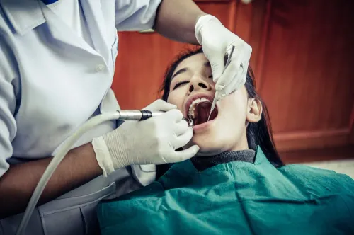
- A surgical handpiece removes only the bone blocking access. The tooth may be divided into sections instead of being forced out whole.
- You feel pressure, vibration, pushing, or pulling, but local anaesthetic should prevent sharp operative pain.
- The socket is cleaned and closed with sutures when needed. Ask whether your quotation includes imaging, the surgical fee, anaesthesia, medicines, follow-up, and treatment of complications.
| Anaesthesia | What you experience | Practical requirements |
|---|---|---|
| Local anaesthesia | You remain awake; the area is numb, but pressure and movement remain noticeable. | Eat and take medicines as instructed unless your clinician says otherwise. |
| Sedation with local anaesthesia | Anxiety and awareness reduce, but sedation does not replace the numbing injection. | Follow fasting instructions and arrange an escort; do not drive afterward. |
| General anaesthesia | You are unconscious during surgery and need closer monitoring. | Fasting, recovery observation, and an adult escort are required; suitability depends on your health and the facility. |
Recovery, pain control, and warning signs after extraction
Pain and swelling commonly worsen during the first 48 to 72 hours after surgical wisdom tooth removal, then gradually improve. Mild bleeding or blood-stained saliva, jaw stiffness, bruising, and difficulty chewing are also expected.
Bite firmly on the gauze for the time advised by your dentist. Use a cold pack on the cheek for short periods, rest with your head raised, and choose soft, cool foods. Avoid smoking, vaping, alcohol, straws, vigorous rinsing, and strenuous exercise because suction or irritation can dislodge the clot.
After 24 hours, clean the other teeth normally and rinse gently with warm salt water if instructed; do not probe the socket.
An NSAID such as ibuprofen, alone or with paracetamol, is a common first-line option when medically suitable. Ask before using an NSAID if you have ulcer disease, kidney disease, take anticoagulants, or are pregnant; check paracetamol limits if you have liver disease or take combination medicines. Do not start antibiotics without advice.
Pain that becomes stronger and throbbing two to five days later, radiates toward the ear, or comes with a foul taste or odour can indicate dry socket. It is delayed healing, not automatically an infection; irrigation, a dressing, and pain control are usual treatments.
Seek urgent dental or medical review for:
- Bleeding that does not stop with firm pressure
- Rapidly increasing facial or neck swelling
- Difficulty breathing or swallowing
- Fever with worsening pain, dehydration, or inability to drink
- New numbness that persists after the anaesthetic should have worn off
Breathing difficulty or expanding neck swelling is an emergency.
How to compare an impacted wisdom tooth dentist in Nashik
A low quotation does not prove a lower-cost procedure. “Extraction” might mean lifting an erupted tooth, or surgical removal involving an incision, bone removal, and sectioning; compare the treatment steps, not just the total.
| Quotation item | Ask the provider to specify | Why it matters |
|---|---|---|
| Assessment and imaging | Consultation, panoramic radiograph, and any CBCT fee | Three-dimensional imaging may be needed when roots approach the inferior alveolar nerve, sinus, or adjacent tooth |
| Procedure | Surgical fee, tooth sectioning, bone removal, and difficulty-related charges | An impacted tooth is not priced like a simple erupted tooth |
| Anaesthesia | Local anaesthesia, sedation, or general anaesthesia, including monitoring | These options involve different preparation, recovery, and fees |
| Aftercare | Medicines, review visits, and management of complications | A quote that excludes follow-up can conceal later costs |
Before agreeing, ask for these points in writing:
- Which tooth is being removed and what the scan shows about root shape, depth, angulation, and nerve contact
- The risk of altered sensation in the lower lip, chin, or tongue for a lower tooth
- For an upper tooth, what happens if an opening into the maxillary sinus develops
- Whether antibiotics are prescribed only for a stated clinical reason
- What emergency contact and review arrangements apply if bleeding, infection, or persistent numbness occurs
When comparing an impacted wisdom tooth dentist in Nashik, ask Dr. Bhonsale's Dental Clinic & Implant Center for the same itemised breakdown. Use identical questions when comparing wisdom tooth extraction in Nashik, because “painless extraction” cannot predict how difficult an impacted tooth will be.
Related service
Tooth Extraction (Including Third Molars) Tooth extraction involves the safe removal of teeth that are damaged, decayed, or impacted. Dr. View service → |
Frequently asked questions
How can you tell whether a wisdom tooth is impacted?
An oral examination and panoramic radiograph show whether the tooth is blocked by its angle, depth, gum, bone, or the neighbouring second molar.
When does an impacted wisdom tooth need removal?
Removal is considered when the tooth causes infection, decay, gum disease, cystic change, damage to the second molar, pain, or another clear problem.
When is monitoring an impacted wisdom tooth safer?
Monitoring can be safer when the tooth has no disease or symptoms, removal carries significant risk, and you can attend planned examinations and imaging.
What happens during wisdom tooth extraction?
The dentist reviews imaging, numbs the area with local anaesthesia, removes gum or bone when needed, sections the tooth if necessary, and places stitches when required.
Which warning signs need attention after extraction?
Contact the dental team for worsening swelling, fever, difficulty swallowing or breathing, uncontrolled bleeding, severe increasing pain, or foul-tasting discharge.
Keywords